2020 FSA Posters
P009: MEDIASTINAL MASS FOLLOWING ORTHOTIC HEART TRANSPLANTATION
Sofia A Lifgren, MD; Adriana S Martini, MD; Jackson Memorial Hospital/UMH
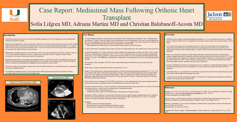
Introduction: Mediastinal masses increase the risks associated with general anesthesia (GA). This can include feared complications as hemodynamic compromise and airway collapse. We present the case of a 52-year-old female, who following a successful orthotic heart transplant with bicaval anastomosis presented with a large mediastinal mass compressing heart structures. The patient had history of nonischemic cardiomyopathy, Ejection Fraction (EF) of 5-10%, NYHA functional classification IV stage D biventricular failure with corresponding severe Mitral Regurgitation and Tricuspid Regurgitation. She presented 20 days after discharge, with complaints of shortness of breath and discharge from sternotomy wound. Imaging demonstrated a mediastinal fluid collection, diagnosed as abscess, displacing her heart and partially compressing her right atrium (RA) and right ventricle (RV). Transthoracic Echocardiography (TTE) on presentation ruled out tamponade and underwent drainage without issue. One month following, she was found to have on computed tomography (CT) of the chest, a large right mediastinal collection with enhancing rim and internal gas with cutaneo-mediastinal fistulous communication suggesting large abscess exerting a significant mass effect on RA, right atrial appendage and moderate mass effect on right superior pulmonary vein and left atrium (LA). It was also noted that there was a leftward and posterior deviation of the superior vena cava (SVC) with moderate narrowing of its lumen as it approaches the RA. Perioperative TTE demonstrated large pericardial effusion. After careful review of imaging and clinical assessment where patient was found to be ventilating well on room air, she was taken to the operating room (OR). A pre-induction radial arterial line (A-line) was placed for continuous hemodynamic monitoring and GA was achieved with standard intravenous induction without incident. Intraoperative transesophageal echocardiography (TEE) was performed to examine fluid collection and aid in surgical planning. Imaging demonstrated a large fluid collection with intraluminal debris adjacent to the RA without tamponade effect measuring 9.9x6.5cm. This was completely drained by the surgical team under our guidance without complications. This case presents an example where careful planning and preparation can ensure safe and successful surgery in a post orthotic heart transplant patient.
Discussion: Mediastinal masses require considerable planning for anesthesia. It is well documented that positive or negative ventilation can alter cardiopulmonary physiology causing collapse placing the patient at risk for death. In this case, the patient’s preoperative studies demonstrated high concern for the possibility of hemodynamic collapse. Imaging demonstrated a large fluid collection compressing the right side of her heart and her SVC. Prior to placing the patient under anesthesia, we performed simple but effective maneuvers to ensure patient could tolerate the transition from negative pressure ventilation to positive pressure ventilation. When creating our anesthetic plan, we also considered the needs of the surgical team which required patient’s relaxation and input from our TEE exam. As anesthesiologist it is our responsibility to both foresee and plan for any adverse events while balancing our goal of providing a safe and effective environment for surgery.

