2020 FSA Posters
P094: IN EACH LOSS THERE IS A GAIN – INCREASED COLOR FLOW GAIN CAN LEAD TO IMAGE ARTIFACTS IN DIALYSIS GRAFTS
Pamela Sharaf, MD; Daniel Perez, MD; Robyn Weisman, MD; University of Miami Health System
Introduction: General anesthesia can be sometimes be avoided during an arteriovenous fistula (AVF) creation or ligation operation by employing a regional anesthesia technique, specifically an infraclavicular or supraclavicular block depending on the location of the fistula and patient-dependent factors. Most patients requiring long-term dialysis and AVFs have associated comorbidities, such as heart failure or obesity, that would make avoiding general anesthesia preferable. The AVF itself can create ultrasound image artifacts due to tissue vibration that requires expertise in image optimization. We will discuss a case report of image artifact during attempted supraclavicular block placement and how to discern true vascularity from artifact.
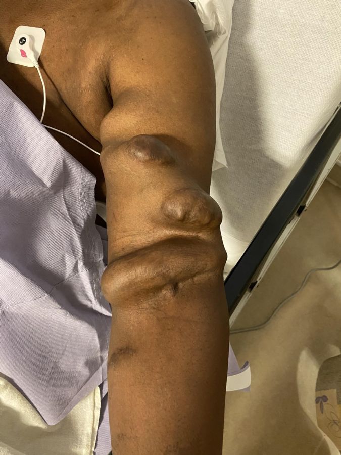
Case Presentation: A 47-year old male was planned to undergo removal of his brachiocephalic venous fistula one-year status post kidney transplantation. Physical examination by his vascular surgeon prior to surgery revealed a “chronically swollen” upper extremity, an “extremely large and aneurysmal” fistula “more than 2 cm in diameter throughout” that “extends from the elbow area to the mid axilla,” “pulsatile but no appreciable thrill,” with a “weak radial pulse”, see Figure 1.
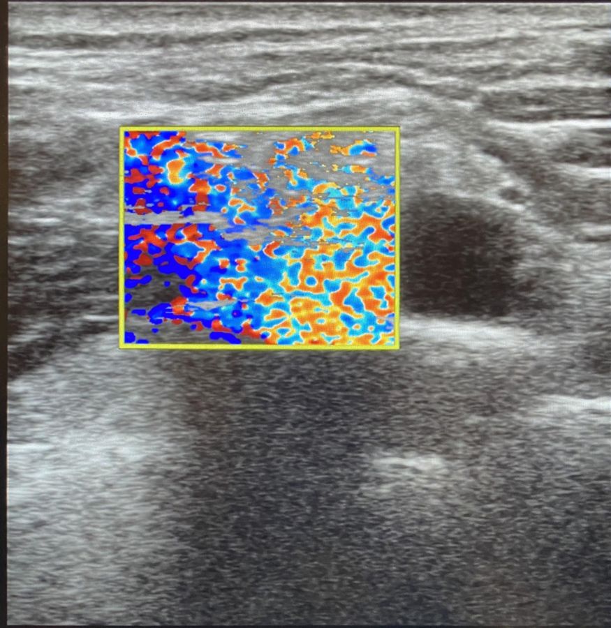
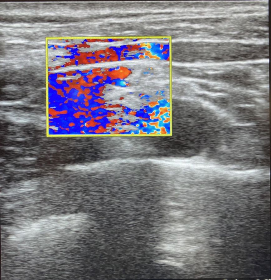
A regional anesthesia technique was planned and supraclavicular brachial plexus block was chosen as it was the farthest point from the fistula that would allow appropriate anesthetic coverage. Upon ultrasound exam using color flow on a Sonosite Xporte, the brachial plexus target seemed to be plagued by increased vascularity surrounding and within the brachial plexus, see Figure 2 & 3. It was decided to switch the anesthetic plan to general anesthesia due to concern for hematoma within the brachial plexus or local anesthetic toxicity (LAST). The patient underwent a successful surgery with an uneventful general anesthetic. Afterwards the images were reviewed by a radiologist, it was determined that the vascular appearance around and in the brachial plexus were artifact as a result of vibration transmitted from the fistula site.
Discussion: Some risks of regional anesthesia include damage to nerves, local infection, and intravascular injection leading to LAST (1). The risks and benefits of block placement must be weighed prior to block placement. Although it was later discovered that the appearance of increased vascular flow within and surrounding the brachial plexus was simply artifact, this affected the decision making process for appropriate risk stratification. Identification of ultrasound artifacts are important skill sets for regionalists. Image optimization can help differentiate artifact versus true increased vascularity.
Some steps that could have been used to minimize artifact on this image would be to decrease the gain on the color. Increased color flow gain settings will lead to “bleeding” to adjacent structures (2). Other strategies for improving image quality are to increase the velocity scale by adjusting the pulse repetition frequency (PRF). This decreases the sensitivity to tissue vibration (3).


1. Jeng CL, Torrillo TM, Rosenblatt MA. Complications of peripheral nerve blocks. Br J Anaesth 2010; 105 Suppl 1:i97.
2. Allan PLP, Baxter GM, Weston MJ. Clinical ultrasound. 3rd ed. Edinburgh: Churchill Livingstone, 2011.
3. Gray AT. Ultrasound-guided regional anesthesia: current state of the art. Anesthesiology 2006; 104:368.
